Orsolya Losonci BSc (Hons), EEBW, Member of the IAABC and IEBWA
MUD FEVER AND RAIN SCALD
Humid, rainy weather, slippery and muddy gateways – a nightmare for all involved. As winter months sneak in so do common equine skin conditions, with conditions such as rain scald and mud fever being prevalent. The causes and treatments for these two conditions are similar, however, their clinical signs and location of these signs differ.
In the acute stage of mud fever, clinical signs typically appear in the form of cracks and lesions at the caudal (back) aspect of the pasterns with fresh pus leaking out of them. Some swelling, redness, irritation, hair loss and crusts may also be present at this stage. In chronic cases, all of the above along with hardened crusts (Figure 1) are commonly present (Hamid, 2016; VeterianKey, 2016; Marsella, 2019; SevernEdge, 2021). Lameness and pain on palpation are typical in these advanced cases and horses may object to treatment. If left untreated, old crusts may stimulate development of further cracks and other skin trauma, eventually leading to heavy scab formation (Hamid, 2016). Mud fever can be a debilitating condition for horses in work and those actively competing. Working in sandy schools or wearing boots may not be appropriate until clinical signs have resolved.

Figure 1. Mud fever at the caudal aspect of the pastern.
Rain scald on the other hand, typically presents in the form of paintbrush-like scabs on the back and the hindquarters of horses (Figure 2). Other clinical signs may include patches of hair loss and dusty-looking hair (Hamid, 2016; Frye et al., 2019).

Figure 2. Characteristic paintbrush like scabs on the horse’s back (Source: Author).
The causative bacterial agents of both conditions include Dermatophilus congolensis, Staphylococcus spp., and Pseudomonas aeruginosa as well as fungi such as Dermatophytes. All of which may be present in the horse’s environment in the form of hyphae (Scott & Miller, 2011) but are harmless in the dormant stage. Even concentrated amounts of these bacteria will not be able to cause infection to an intact skin (Scott & Miller, 2011), which is why some horses may live in wet, muddy conditions for a prolonged period of time without developing these conditions.
Certain types of soil and old crusts may act as a temporary reservoir for Dermatophilus c. (Scott & Miller, 2011; Aufox et al., 2018; Marsella, 2019) where the bacteria remain dormant until environmental conditions become ideal for reactivation (VeterianKey, 2016). Bacteria tend to thrive in a wet, warm environment, such as the caudal aspect of the pastern. They enter the epidermis (outer layer of the skin) (Figure 3) at the site of an injury which could be an open wound or an insect bite, and trigger an inflammatory response, causing scabs or crust formation at the site of trauma (VeterianKey, 2016; Aufox et al., 2018; Marsella, 2019; SevernEdge, 2021). Transmission of the causative agents is possible via biting insects and potentially also via direct contact with acute lesions (Underwood et al., 2015). The incubation period is extremely variable, lasting anywhere between one and 34 days (Scott & Miller, 2011).

Figure 3. Layers of the skin that form a protective barrier between the horse and its environment.
PREDISPOSING FACTORS
In wet and muddy conditions, the equine skin may remain damp for a prolonged period. Moisture causes the release of infective zoospores of bacteria or fungi into the outermost layer of the skin, the epidermis (Cullinane et al., 2006; Marsella, 2019). At the same time, protective bacteria on the skin such as Bacillus spp. and Staphylococcus spp. are diluted due to prolonged exposure to heat and humidity (Hamid, 2016; Frye et al., 2019). Damage to the oily layer of the skin may also happen (Ashton, 2020), and imbalances in these epidermal constituents may play a role in the development of infection, although thickness of the skin was not found to be a contributing factor (Scott & Miller, 2011). According to Scott & Miller (2011) the intensity of rainfall may be more important than the total amount of rain, suggesting that a sudden insult to the skin also increases the risk of these conditions.
The immune response of the horse is critical in determining an individual animal’s resistance to infection (Marsella, 2019). Horses with a weakened immune system such as those with Pituitary Pars Intermedia Dysfunction (PPID – commonly referred to as Cushings) may require nutritional support to aid resistance against bacterial skin conditions.
Usually, both skin conditions develop from the late autumn until early spring months, with the first lesions tending to occur 2–3 weeks after the onset of the rainy season. The likelihood of actual onset is heavily dependent on management practices (Marsella, 2019) (Table 1).

Table 1.
MANAGEMENT CONSIDERATIONS
Prevention is always better than cure and is less stressful for all involved, with good management practices being of primary importance. Flexibility to adjust routines and implement changes to the horse’s management are essential for prevention during wet weather and successful treatment should the conditions develop.
Feral (free-living) and domestic horses alike naturally seek to avoid muddy areas. However, in domestic situations this is often not possible due to restricted amounts of space and motivating factors to stay in busy areas such as the gate, the hay pile or the shelter. This motivation may be greater than the urge to move away to a drier area, leading to horses being exposed to mud and rain for prolonged periods of time, and an increased risk of mud fever and rain scald developing. However, we all know that mud is an unavoidable part of owning and working with horses. A small amount of mud may even be beneficial to the horse’s skin, helping to keep parasites away and stimulating the sebaceous gland to produce natural oils that act as a barrier against dust, water and bacteria (SevernEdge, 2021) and which are beneficial for optimal hoof function. Exposure to too much deep mud, however, can be detrimental to your horse’s health.
HOW MUCH MUD IS TOO MUCH?
Generally speaking, if the mud covers the hooves and reaches the coronary band it is considered to be suboptimal. This level of mud can increase the chances of mud fever, loosing shoes and soft tissue injuries, just to name a few. Ideally the gate should be at the highest point of your field to avoid the build-up of mud, although such a busy area may still become muddy. To limit exposure to mud, the following options should be considered:
- Move the horse to another field.
- Rotate fields to avoid overuse.
- Block off muddy areas with electric fence.
- Put down mats or wood chip as an alternative footing.
- Moving gates, hay and other commonly used areas.
- Stabling the horse overnight or alternatively, keeping the horse in.
- Placing the hay in several piles encourages horses to move around instead of eating in one place. This also mimics natural browsing behaviour and correct head and neck position - always make sure there are more hay piles than horses present in the field to avoid frustration and conflict amongst the herd.
While daily turnout is crucial for equine welfare, limiting the skin's exposure to mud and moisture may be a greater, short-term concern, particularly for severe cases. Options include individual stabling or hard standing areas that still allow free movement and time outside with other horses. If stabled, the stable and the horse’s legs should be kept clean and dry at all times. Picking up droppings several times during the day and daily removal of soiled bedding is the most appropriate course of action for mud fever cases. To help keep your horse relaxed when stabled seek out enrichment options, stable toys and activities you can do to keep them occupied, along with providing enough forage to allow trickle feeding behaviour.
Bedding may also play a primary role in managing this condition. According to Yarnell et al., (2017) pine bedding (shavings) (Figure 4) has been shown to discourage or slow down bacterial growth of equine disease-causing bacteria including Dermatophilus c., one of the bacteria responsible for mud fever and rain scald. Compared to other alternatives, such as straw, pine shavings showed the largest moisture-holding capacity of all bedding types tested, making it an appropriate choice for mud fever cases where moisture is to be avoided at all times.

Figure 4. A clean stable helps to manage and reduce the likelihood of mud fever.
According to Kim & Shin (2005) and Zeng et al. (2012) foot and respiratory health of horses improved when horses were stabled on pine-based products. The antimicrobial properties of pine could be associated with stilbenes, a polyphenol found in the wood of pinus species (Välimaa et al., 2007), especially corsican pine (pinus nigra). Choice of bedding could therefore be an important factor in the appropriate management of the condition.
If your horse is kept at a yard where you cannot influence their management, there are still a few things you could do to help prevent mud fever and rain scald developing:
USE TURNOUT BOOTS
Turnout boots are developed to keep the legs clean and dry while your horse is in the field. Look for those that provide all-round protection, made from a water-repellent yet breathable material. Also, ensure that they are correctly fitted to avoid slipping or rubbing of the skin. Ideally, you would have two sets so that you can clean and dry them for the next morning.
CLIPPING LONG FEATHERS
Clipping allows easy monitoring of the affected area, facilitates treatment, and allows more air to the lesions. Avoid clipping your horse’s feathers without a valid reason. Horses’ coats are designed to guide water droplets to the ground without them coming in contact with the skin. Clipping off feathers could disrupt this natural mechanism and could in fact increase the risk of mud fever.
AVOID FREQUENT WASHING OF LEGS
Washing the legs on a daily basis removes the protective oils in the skin and may increase susceptibility to mud fever (SevernEdge, 2021). If you do need to wash your horse’s legs, ensure that they are dried thoroughly with clean towels after washing, and particularly before exercise or turning the horse back out. A warm and moist environment will encourage the bacteria to grow.
Sometimes a better way to keep the lower leg clean is to provide a thick bed in the horse’s stable to absorb the moisture, wait for the mud to dry overnight, and remove the dry mud the next day. Use a gentle brush with soft bristles to avoid causing microtrauma to the skin. This is also the perfect time to check for signs of mud fever, such as new crusts.

Table 2.
TREATMENT
Once present, bacteria are difficult to eliminate. They were the first organism on Earth and their adaptive nature enabled them to still be present today. They can even adapt to antibiotic treatment and produce up to 16 million offspring per day (McDowell & Rowling, 2003).
Research on Dermatophilus c., one of the bacteria responsible for rain scald and mud fever, found that it can remain dormant in old crusts at temperatures of 28-31°C for over 3.5 years, even when heated and dried at 100°C, and for 13 years when frozen (Pilsworth & Knottenbelt, 2007; Scott & Miller, 2011). Favourable conditions for its growth include certain types of soil and moisture content, although the pH of the soil was found to not affect its survival (Scott & Miller, 2011). Such findings demonstrate the challenge of controlling such bacteria and treating these common skin conditions.

Figure 5. Horses with an underdeveloped topline can be prone to rain scald (Source: Author).
As rain scald and mud fever are multifactorial conditions, the same treatment plan may not be appropriate for each horse. First and foremost, a proper diagnosis is essential as rain scald may be mistaken for ringworm, a highly contagious fungal infection, and mud fever can be confused with pastern and cannon leukocytoclastic vasculitis - an allergic reaction to sunlight and other environmental factors (SevernEdge, 2021).
Underlying conditions such as fungal or mite infections, allergies, or a suppressed immune system, can also influence treatment options (SevernEdge, 2021). Vets recommend treatment plans on a case-by-case basis after evaluating the severity of the individual case and any underlying primary causes. Treatment depends on how advanced the case is and typically involves topical or systemic antibiotics (Hamid, 2016), anti-inflammatories and analgesics. Prognosis is usually very good for average cases where the condition resolves in just a few weeks. However, treatment may be prolonged for horses with a weakened immune system (Marsella, 2019).
Make sure to notify your vet if the condition does not improve or becomes worse. Documentation of skin lesions via regular photographs could be an objective way of tracking the healing process. Removal and proper disposal of crusts may be advised for preventing reinfection (VeterianKey, 2016), although you should discuss with your vet if the removal of crusts is appropriate in your horse’s case, and the most appropriate method to perform such removal to avoid damaging the skin further. Scab removal in rain scald cases may also be required. Pick paintbrush-like scabs with your fingertips but avoid picking out those that do not come off easily. Leave them for a few days until they are ready to be removed. Regardless of treatment method, wearing disposable gloves when administering treatment is advised as the condition may spread to humans, with immunosuppressed individuals being more at risk (Marsella, 2019).
When antifungal and antibacterial scrub is recommended, apply it gently to avoid microtrauma to the skin. It is generally applied on a three- to four-day basis to prevent dry skin. After application, rinse and dry the skin thoroughly. Application of an antibacterial barrier cream may also be recommended before turnout, however, always make sure that the area is dry before applying and avoid product buildup.
Horses that object to treatment procedures could benefit from the use of a lick or chaff as a distraction, or operant conditioning. Ultimately, restraint or sedation may be necessary.
HERBAL REMEDIES
Herbal remedies may be used as complementary treatment options. Anti-inflammatory herbs cleanse the area to aid the inflammatory process rather than suppressing it. Such herbs could be particularly helpful in inflammatory skin conditions (Self, 2005) (Table 3).

Table 3. Herbal remedies and their effects.
Colloidal silver may also help in acute or chronic infected wound management as it is known for its antibacterial, antiviral and antifungal effects (McDowell & Rowling, 2003; Dharmshaktu et al., 2016). However, according to Schwarcz, (2019) colloidal silver may cause argyria, a condition where silver particles deposit in the skin, causing it to turn grey, making this a consideration for dark coloured horses or those with a showing career. Natural herbal creams may also be applied where appropriate. Look for ones with lavender, which acts as a natural insect repellent.
The dosage of herbal remedies as well as possible adverse health effects are not always clear. For appropriate application and dosage, consult with your vet and a qualified equine herbalist or nutritionist.
NUTRITIONAL CONSIDERATIONS
A healthy skin protects the body from harmful environmental factors such as weather changes and injury. A balanced diet with optimum levels of Zinc, Copper and Biotin will support skin health from within. Quality protein is also an essential requirement as 80-85% of hair is composed of protein. Supplementation of antioxidants to the diet in the form of Vitamins C and E can also help “mop up” free radicals in the body and ease itchy, irritated skin. Furthermore, Omega-3 fatty acids are known for their anti-inflammatory properties (Geor et al., 2013), with a correct ratio of Omega-3:Omega-6 fatty acids being highly beneficial for skin conditions, especially in acute or chronic inflammatory cases (Geor et al., 2013). For an in-depth explanation of Omega-3 and -6 fatty acids, including how they support the skin, please refer to Essential fatty acids by Dr. Stephanie Wood.
SUMMARY
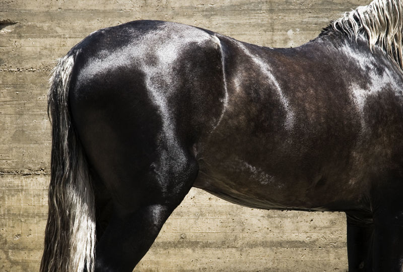
In conclusion, mud fever and rain scald are both multifactorial equine skin conditions. Prolonged exposure to wet environments (Figure 6) and skin trauma are the two main predisposing factors. Prognosis for average cases is good with a relatively simple course of treatment, however, owners should aim to prevent the issue where possible. Eliminating the primary, underlying cause will usually result in a quick recovery and ideally, no recurrence of the condition.
REFERENCES
Ashton, S. (2020). How to Treat Mud Fever. EverythingHorse. [online]. Available from: https://everythinghorseuk.co.uk/how-to-treat-mud-fever/ [Accessed November 22, 2021].
Aufox, E., Frank, L., May, E., & Kania, S. (2018). The prevalence of Dermatophilus congolensis in horses with pastern dermatitis using PCR to diagnose infection in a population of horses in southern USA. Veterinary Dermatology, 29(5): 425-e144.
Cullinane, A., Barr, B., Bernard, W., Duncan, J., Mulcahy, G., Smith, I., & Timoney, J. (2006). The Equine Manual. 2nd ed. Davis CA: Elsevier.
Dharmshaktu, G., Singhal, A., & Pangtey, T. (2016). Colloidal silver-based nanogel as nonocclusive dressing for multiple superficial pellet wounds. Journal of Family Medicine and Primary Care, 5(1): 175–177
Frye, C., Bei, D., Parman, J., Adam, J., Houlihan, J., & Rumore, A. (2019). Efficacy of Tea Tree Oil in the Treatment of Equine Streptothricosis. Journal of Equine Veterinary Science, 79(1): 79-85.
Geor, R., Harris, P., & Coenen, M. (2013) Equine Applied and Clinical Nutrition. 1st ed. Edinburgh: Elsevier.
Hamid, M. (2016). Skin Diseases of Cattle in the Tropics. 1st ed. London: Academic Press.
Kim, Y., & Shin, D. (2005). Volatile components and antibacterial effects of pine needle (Pinus densiflora S. and Z.) extracts. Food Microbiology, 22(1): 37-45.
Knottenbelt, D. (2012). The Approach to the Equine Dermatology Case in Practice. Veterinary Clinics of North America: Equine Practice, 28(1): 131-153.
Marsella, R. (2019). Manual of Equine Dermatology. 1st ed. Oxfordshire: CAB International.
McDowell, R., & Rowling, D. (2003). Herbal Horsekeeping. 1st ed. London: J. A. Allen.
Nicholson, W., Sonenshine, D., Noden, B., & Brown, R. (2019). Medical and Veterinary Entomology. 3rd ed. Starkville MS: Academic Press.
Pilsworth, R., & Knottenbelt, D. (2007). Skin Diseases Refresher. American Association of Equine Practitioners.
Schwarcz, J. (2019). Does Taking “Colloidal Silver” Have Health Benefits?. Mc. Gill. [online]. Available from: https://www.mcgill.ca/oss/article/health-you-asked/does-taking-colloidal-silver-have-health-benefits [Accessed November 22, 2021].
Scott, D., & Miller, W. (2011). Equine Dermatology. 2nd ed. Ithaca NY: Elsevier.
Self, H. (2005). Veteran Horse Herbal. 1st ed. Buckingham: Kenilworth Press.
SevernEdge (2021). Under The Skin: The Truth About Mud Fever. Severn Edge Vets. [online]. Available from: https://www.severnedgevets.co.uk/equine/advice/under-skin-truth-about-mud-fever [Accessed November 22, 2021].
Underwood, W., Blauwiekel, R., Delano, M., Gillesby, R., Mischler, S., & Schoell, A. (2015). Chapter 15 - Biology and Diseases of Ruminants (Sheep, Goats, and Cattle). In Laboratory Animal Medicine. London: Academic Press.
Varró, A. (2006) Gyógynövények gyógyhatásai. 1st ed. Debrecen: Black & White Könyvkereskedés Kft.
Välimaa, A., Honkalampi-Hämäläinen, U., Pietarinen, S., Willför, S., Holmbom, B., & Wright, A. (2007). Antimicrobial and cytotoxic knotwood extracts and related pure compounds and their effects on food-associated microorganisms. International Journal of Food Microbiology. 115(2): 235-243.
VeterianKey (2016). Dermatophilosis. Veterian Key. [online]. Available from: https://veteriankey.com/dermatophilosis/ [Accessed November 22, 2021].
Yarnell, K., Le Bon, M., Turton, N., Savova, M., McGlennon, A., & Forsythe, S. (2017). Reducing exposure to pathogens in the horse: a preliminary study into the survival of bacteria on a range of equine bedding types. Journal of Applied Microbiology, 122(1): 23-29.
Zeng, W., He, Q., Sun, Q., Zhong, K., & Gao, H. (2012). Antibacterial activity of water-soluble extract from pine needles of Cedrus deodara. International Journal of Food Microbiology, 153(1-2): 78-84.


